
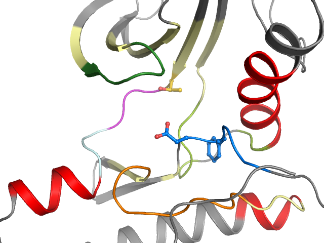
3P1A - chain A (model B) | Protein kinase, membrane associated tyrosine/threonine 1
Structure information
| PDB: | 3P1A |
| PubMed: | - |
| Release date: | 2010-11-03 |
| Resolution: | 1.7 Å |
| Kinase: | PKMYT1 (MYT1) |
| Family: | WEE |
| Group: | Other |
| Species: | HUMAN |
| Quality Score: | 8 |
| Missing Residues: | 0 |
| Missing Atoms: | 0 |
| DFG conformation: | in |
| αC-helix conformation: | in |
| Salt bridge KIII.17 and EαC.24: | Yes (2.9Å) |
| ASP rotation (xDFG.81) : | 351° |
| PHE rotation (xDFG.82) : | 356° |
| Activation loop position: | -3.1Å |
| αC-helix position: | 17.9Å |
| G-rich loop angle: | 58.2° |
| G-rich loop distance: | 18.1Å |
| G-rich loop rotation: | 53° |
Other models from this PDB:
2D & 3D views
Binding pocket waters
The following waters were found in the defined clusters:
I2
H-bond protein
I3
H-bond protein
I4
H-bond protein
I5
H-bond protein
I7
No H-bonds
I9
No H-bonds
Binding pocket sequence
| Uniprot | SRLGHGSYGEVFKYAVKRSRKLAEVGSHEKVGPCCVRLEQAYLQTELC-GPSLQQHCEAHLHSQGLVHLDVKPANIFLLGDFGLL |
| Structure: | SRLGHGSYGEVFKYAVKRSRKLAEVGSHEKVGPCCVRLEQAYLQTELC_GPSLQQHCEAHLHSQGLVHLDVKPANIFLLGDFGLL |
Modified residues
No modified residues identified.