
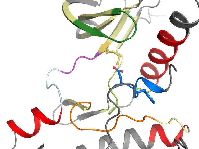
3CC6 - chain A (model B) | Protein tyrosine kinase 2 beta
Structure information
| PDB: | 3CC6 |
| PubMed: | - |
| Release date: | 2008-03-11 |
| Resolution: | 1.6 Å |
| Kinase: | PTK2B (PYK2) |
| Family: | FAK |
| Group: | TK |
| Species: | HUMAN |
| Quality Score: | 8 |
| Missing Residues: | 0 |
| Missing Atoms: | 0 |
| DFG conformation: | in |
| αC-helix conformation: | in |
| Salt bridge KIII.17 and EαC.24: | Yes (3.5Å) |
| ASP rotation (xDFG.81) : | 78° |
| PHE rotation (xDFG.82) : | 4° |
| Activation loop position: | -4.8Å |
| αC-helix position: | 18.4Å |
| G-rich loop angle: | 69.4° |
| G-rich loop distance: | 19.8Å |
| G-rich loop rotation: | 66.5° |
Other models from this PDB:
2D & 3D views
Binding pocket waters
The following waters were found in the defined clusters:
I1
H-bond protein
I3
No H-bonds
I6
No H-bonds
I9
No H-bonds
Binding pocket sequence
| Uniprot | RILGEGFFGEVYEVAVKTCKFMSEAVIMKNLDPHIVKLIGIWIIMELYPYGELGHYLERYLESINCVHRDIAVRNILVLGDFGLS |
| Structure: | RILGEGFFGEVYEVAVKTCKFMSEAVIMKNLDPHIVKLIGIWIIMELYPYGELGHYLERYLESINCVHRDIAVRNILVLGDFGLS |
Modified residues
No modified residues identified.