
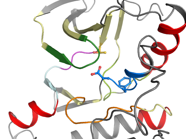
2G1T - chain C | ABL proto-oncogene 1, non-receptor tyrosine kinase
Structure information
| PDB: | 2G1T |
| PubMed: | 16640460 |
| Release date: | 2006-05-23 |
| Resolution: | 1.8 Å |
| Kinase: | ABL1 |
| Family: | Abl |
| Group: | TK |
| Species: | HUMAN |
| Quality Score: | 8 |
| Missing Residues: | 0 |
| Missing Atoms: | 0 |
| DFG conformation: | in |
| αC-helix conformation: | out |
| Salt bridge KIII.17 and EαC.24: | No (14.7Å) |
| ASP rotation (xDFG.81) : | 310° |
| PHE rotation (xDFG.82) : | 311° |
| Activation loop position: | -5Å |
| αC-helix position: | 21.8Å |
| G-rich loop angle: | 50.2° |
| G-rich loop distance: | 14.8Å |
| G-rich loop rotation: | 53.1° |
Other models from this PDB:
2D & 3D views
Binding pocket waters
The following waters were found in the defined clusters:
I3
H-bond protein
I4
H-bond protein
I5
H-bond protein
I6
H-bond protein
I11
H-bond protein
Binding pocket sequence
| Uniprot | HKLGGGQYGEVYEVAVKTLEFLKEAAVMKEIKPNLVQLLGVYIITEFMTYGNLLDYLREYLEKKNFIHRDLAARNCLVVADFGLS |
| Structure: | HKLGGGQYGEVYEVAVKTLEFLKEAAVMKEIKPNLVQLLGVYIITEFMTYGNLLDYLREYLEKKNFIHRDLAARNCLVVADFGLS |
Modified residues
No modified residues identified.