
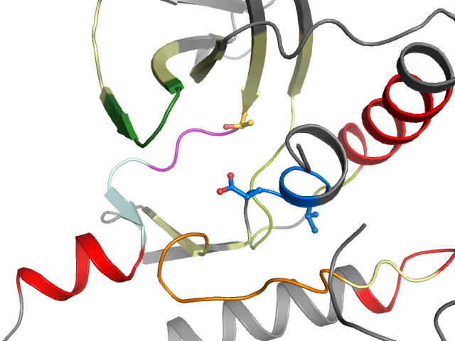
5O1V - chain A (model B) | WNK lysine deficient protein kinase 3
Structure information
| PDB: | 5O1V |
| PubMed: | - |
| Release date: | 2017-06-28 |
| Resolution: | 1.72 Å |
| Kinase: | WNK3 (Wnk3) |
| Family: | WNK |
| Group: | Other |
| Species: | HUMAN |
| Quality Score: | 9 |
| Missing Residues: | 0 |
| Missing Atoms: | 10 |
| DFG conformation: | in |
| αC-helix conformation: | out |
| Salt bridge KIII.17 and EαC.24: | No |
| ASP rotation (xDFG.81) : | 321° |
| PHE rotation (xDFG.82) : | 15° |
| Activation loop position: | -3.6Å |
| αC-helix position: | 20.6Å |
| G-rich loop angle: | 56.9° |
| G-rich loop distance: | 16.2Å |
| G-rich loop rotation: | 47° |
Other models from this PDB:
2D & 3D views
Binding pocket waters
The following waters were found in the defined clusters:
I1
H-bond protein
I3
H-bond protein
I4
H-bond protein
I5
H-bond protein
I7
No H-bonds
Binding pocket sequence
| Uniprot | IELGRGAFKTVYKVAWCELRFKEEAEMLKGLQPNIVRFYDSVLVTELMTSGTLKTYLKRHTRTPPIIHRDLKCDNIFIIGDLGLA |
| Structure: | IELGRGAFKTVYKVAWCELRFKEEAEMLKGLQPNIVRFYDSVLVTELMTSGTLKTYLKRHTRTPPIIHRDLKCDNIFIIGDLGLA |
Modified residues
Residue 304 (not in pocket)
Phosphorylated serine